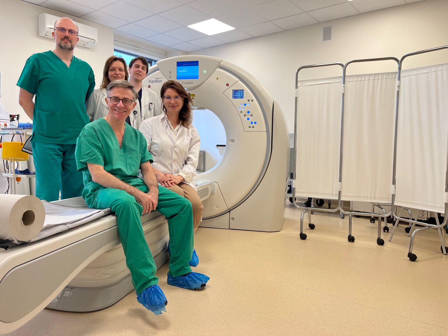
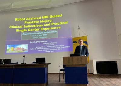
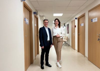
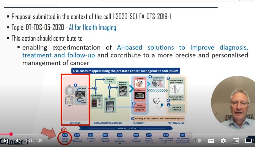
On May 15-16, the National Cancer Institute (NVI) held an international scientific practical conference “Local prostate cancer diagnosis”. The event was organized by the Department of Diagnostic and Interventional Radiology of NVI with guest lecturer prof. Joan C. Vilanova from Girona Clinic, ProCancer-I project partner (Spain), Lithuanian Urological Society and European Council of Urology certified Prostate Cancer Treatment Competence Center -Oncourology Department.
“NCI has been operating a prostate cancer diagnostic and treatment competence center certified by the European Council of Urologists for the sixth year – the only one of its kind in the Baltic States. Urological centers, where prostate cancer is diagnosed and treated using the most advanced and latest methods and possibilities, are accredited by the European Council of Urologists. A prerequisite is that such a center combines clinical work with research. And when it comes to prostate cancer diagnosis, accurate radiological imaging tests and other necessary invasive tests allow for personalized prostate cancer treatment for our patients. The two-day scientific practical conference made it possible to share experience, discuss, and practically demonstrate the new methodologies,” says NCI deputy director for the clinic, Dr. Marius Kinčius.
“NCI participapates at ProCancer-I project, dedicated to artificial intelligence tools that facilitate the diagnosis of prostate cancer. During this project, we got to know professor Joan C. Vilanova of the University of Girona in Catalonia, who is both the author of books on pelvic and prostate imaging and a guest lecturer in magnetic resonance imaging training. Thanks to the ProCancer-I project, we not only cooperate and share experience with prostate cancer reference centers and profesionals, but we can also acquire tools for robotic biopsy,” says Dr. Jurgita Ušinskienė.
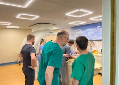
The conference consisted of presentations by NCI doctors radiologists and oncourologists, a guest from Spain, and a practical part – targeted prostate biopsies performed under the direct control of a magnetic resonance (MRI) navigation robotic system, as well as ultrasound fusion of MRI images.
“Prof. Joan C. Vilanova was the first in Spain to introduce a robotic biopsy system under MRI control. This practice began to be applied not so long ago, in 2019. The professor performs over 150 such biopsies a year,” says radiologist dr. Rūta Briedienė.
“The European guidelines are very clear: if a patient has elevated PSA, prostate cancer can be suspected. However, the main method by which we can see prostate cancer is MRI. So if a patient is suspected of having prostate cancer, we do an MRI. If we see a suspicious focus on MRI, we need to evaluate whether it is malignant. We take a biopsy for that. Today, the best option is to take a biopsy directly from the focus. Biopsy under MRI control, using a robotic system, is performed under local anesthesia, takes only a dozen minutes, is convenient for the patient, uncomplicated for the performing staff, and urologists receive an accurate answer as to whether the patient has clinically significant prostate cancer,” shares Prof. Joan C. Vilanova.
According to the professor, since prostate biopsy under MRI control is a relatively new diagnostic procedure, it is very important for radiologists from different countries to share their experience with each other.
“Such diagnostic procedures are relatively new. Not many cancer centers do them. Therefore, it is very important that radiologists from different countries share their experiences and learn from each other,” says prof. Joan C. Vilanova.
The conference was also attended by a guest from Klaipėda – Artūras Čiuvašovas, the head of the Radiology and Tomography Departments of the Klaipėda Republican Hospital, who was the first in the Nordic and Baltic countries and Poland, to perform prostate biopsy under MRI control.
“What we are currently seeing in the diagnosis of prostate cancer is a big change that has come with MRI – not only with the ability to see the cancer during the examination, but also with the new challenge of how to diagnose it, already seeing the tumor.” An MRI-guided biopsy allows us to see the cancer in real time, take a biopsy from it, and examine it. Therefore, we see this procedure becoming more popular. We use a robotic system that automates our routine work, speeds up the procedure, and makes it available in everyday practice. A decade ago, MRI control of biopsies was a 2-hour, skill- and time-consuming work at research centers. The robotic system simplifies this – half an hour is enough to perform the examination. The demand for this procedure is very high. It is appreciated by patients and urologists. At the Klaipėda Republican Hospital, we have performed more than 500 tests in this area and we see a growing demand for them. Which is likely to increase in the future. It is a great honor for us to gain experience at NVI, which is the leader in oncology in Lithuania,” A. Čiuvašovas shares his thoughts.

