The Quantitative Translational Imaging in Medicine (QTIM) Lab (https://qtim-lab.github.io) at the Athinoula A. Martinos Center for Biomedical Imaging (https://www.martinos.org) is a collaborative and interdisciplinary group of physicians and engineers/computer scientists working at the forefront of the application of machine learning and artificial intelligence to the clinical interpretation of medical images. As part of the Massachusetts General Hospital, we are well situated to access the collaborations and data we need to address challenges related to the direct use of AI in medicine to improve patient care. In our group, we have a history of publications in high-impact journals, and our alumni have a track record of independent faculty positions and impactful positions in industry. We are dedicated to providing a supportive and flexible working environment for our team, and mentorship at all levels of career development.
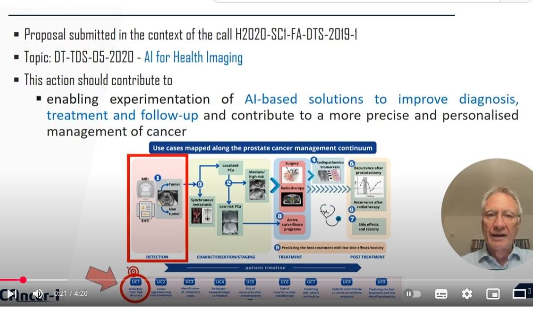
We are seeking a post-doctoral research fellow to work on the application of deep learning to medical image analysis. You will conduct original research aligned with the laboratory’s research agenda but with significant scope for self-directed research. The main focus of this position will be on the application of deep learning to diagnose, characterize, and prognosticate prostate cancer in multi-parametric magnetic resonance imaging (MRI) to produce clinically-actionable insights while addressing technical challenges including lack of interpretability and learning from distributed data sources. You will be working as part of an international consortium (ProCAncer-I) with a large dataset of imaging studies from multiple sites and vendors. In this role, you will work in alongside computer scientists, medical physicists, and physician-scientists at the Massachusetts General Hospital (MGH) in Boston, Massachusetts.
The primary responsibilities of this position will be:
– Conduct research projects in the area of medical image analysis with deep learning, with a primary focus on prostate cancer MRI.
– Assist graduate students, medical students and interns in their research projects.
– Prepare articles and abstracts for high impact scientific journals and conferences.
– Propose and execute novel research projects, in preparation for developing an independent research agenda within the field of medical image analysis.
– Assist with the preparation of applications for research grants.
You must have:
– A PhD in a field related to medical image analysis, such as computer science, engineering, applied mathematics, or physics.
– Expertise in state-of-the-art deep learning.
– Previous experience working in at least one area of image analysis.
For further Information, please contact :Christopher Bridge (cbridge@mgh.harvhard.edu) with questions.


