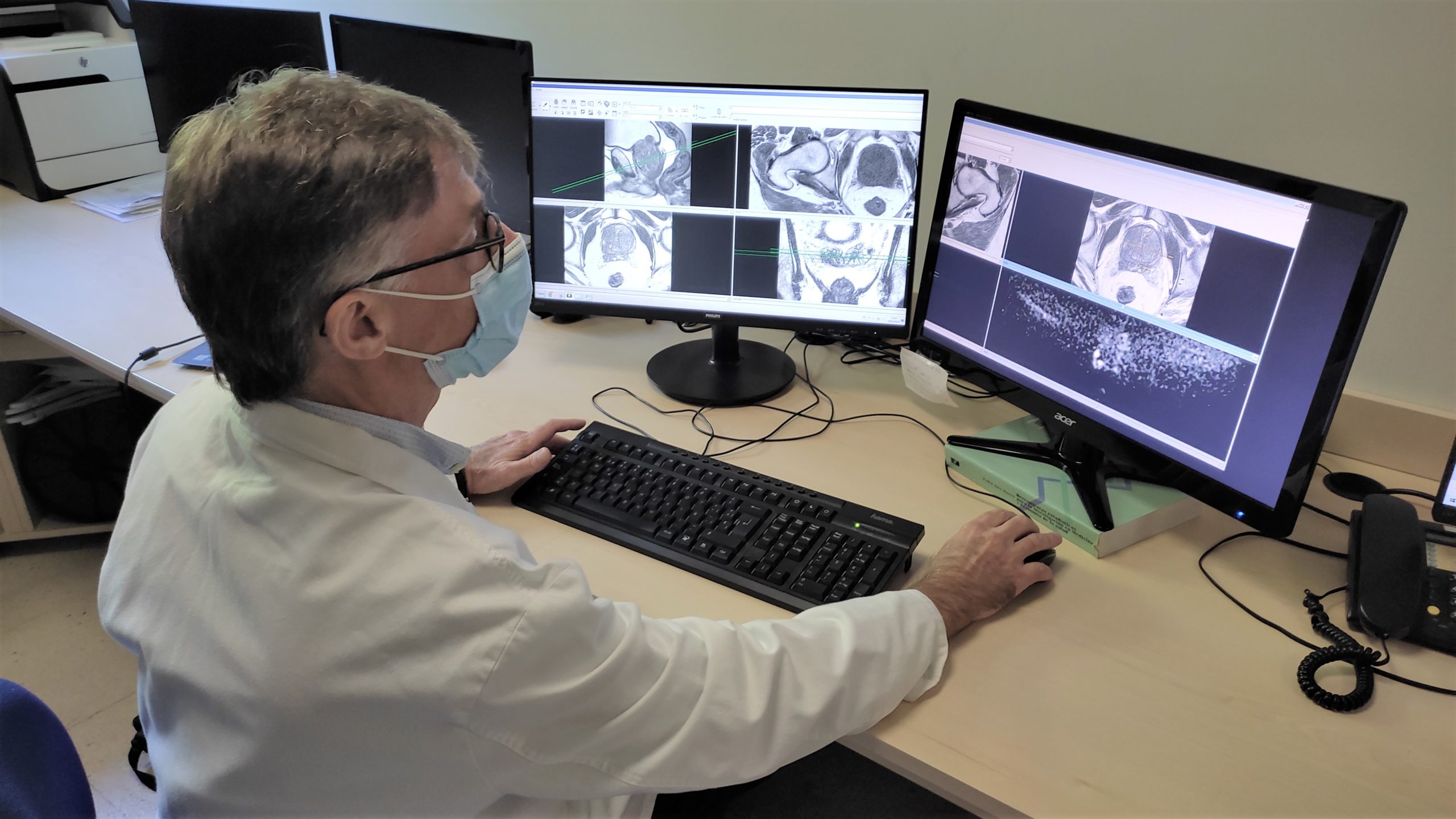
Interview with Dr. Kai Vilanova, Director MRI Clínica Girona, Coordinator Radiology, Faculty of Medicine, University of Girona. Director School of MRI-ESMRMB (IDIBGI)
How can the results of the ProCAncer-I project help radiologists?
What are the problems that professionals encounter today in diagnosing, treating and monitoring prostate cancer, does this project want to solve?
Prostate cancer is currently diagnosed with an MRI scan. MRI consists of the acquisition of different images that provide morphological and physiological information of the prostate. The MRI scan provides about 500-1000 images that the radiologist has to analyze to provide a final report of the degree of suspicion of having prostate cancer. This analysis of each scan involves identifying many parameters manually. The possibility of having software that allows all the necessary parameters in an MRI scan to be identified and evaluated automatically would be a great advantage for the radiologist’s work. This is precisely one of the main objectives of the project; to develop intelligent analysis software to aid in the diagnosis of prostate cancer.
What advantages will this tool have for clinical practice?
The possibility of having an intelligent prostate MRI software means a great saving in analysis time, but also the most important is to improve the diagnostic accuracy to better detect cancer. These tools can detect and classify suspicious prostate cancer lesions, especially useful in cases where lesions are very small or difficult to detect with the naked eye. The artificial intelligence software can automatically segment the prostate, which saves time and improves accuracy by delineating the contours of the prostate gland. It also allows automatic report generation, which helps standardize and streamline the reporting process for the radiologist. Artificial intelligence can analyze the MRI results and generate a report containing relevant findings and recommendations for the patient. The technological tools developed by the project will improve the results of prostate MRI analysis performed by radiologists at all stages of cancer; from diagnosis, staging, prognosis, treatment and avoiding cancer recurrence. In addition, these technological tools that the ProCAncer-I project has developed, give us healthcare personnel the opportunity to free up part of our working day, dedicated to healthcare visits, and use it to continue with research to improve the health of our patients.
What are the challenges, and how will you overcome them?
The main difficulties in the development of these tools are at different levels. Firstly, the difficulty in recruiting the large amount of MRI data and clinical data from more than 15.000 patients and also the need to anonymize all this data. On the other hand, one of the main challenges of these intelligent software programs is the difficulty in being able to integrate into all the computer systems of the different hospitals. These tools may work when they are developed in the laboratory, but in real clinical practice they may not work properly. Therefore, one of the difficulties is to be able to validate these intelligent software programs for their real utility in any hospital. Although their adaptation and operation involves some difficulty, the fact of incorporating radiologists like us into the international consortium of ProCAncer_I also allow us to include our perspective of how the hospital works and take it into account from the very beginning of the design of the tool.
In short, we are sure that the advantages of this new system will make it worthwhile to take this next step of integration in the future, so that the public can benefit from the results of the project.

Dr. Kai Vilanova

